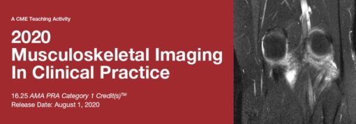
2020 Musculoskeletal Imaging In Clinical Practice (CME VIDEOS)
Musculoskeletal Imaging in Clinical Practice is a review of clinical applications concerning the diagnosis, treatment and management of the musculoskeletal disorders. Throughout the activity modern imaging strategies, surgical correlation and the need for intra-disciplinary teamwork when deciding the most effective patient management is addressed. Faculty share techniques and tips in image interpretation of musculoskeletal injuries and pathology.
This CME activity is designed to educate diagnostic imaging physicians who supervise and interpret musculoskeletal MRI. In addition, referring physicians who order musculoskeletal MRI should gain an appreciation of the strengths and limitations of these types of procedures.
Educational Symposia
Physicians: Educational Symposia is accredited by the Accreditation Council for Continuing Medical Education (ACCME) to provide continuing medical education for physicians.
Educational Symposia designates this enduring material for a maximum of 16.25 AMA PRA Category 1 Credit(s)TM. Physicians should claim only the credit commensurate with the extent of their participation in the activity.
SA-CME: Credits awarded for this enduring activity are designated “SA-CME” by the American Board of Radiology (ABR) and qualify toward fulfilling requirements for Maintenance of Certification (MOC) Part II: Lifelong Learning and Self-assessment.
All activity participants are required to take a written or online test in order to be awarded credit. All course participants will also have the opportunity to critically evaluate the program as it relates to practice relevance and educational objectives.
AMA PRA Category 1 Credit(s)TM for these programs may be claimed until July 31, 2023.
This CME activity was planned and produced by Educational Symposia, the leader in diagnostic imaging education since 1975.
This CME activity was planned and produced in accordance with the ACCME Essential Areas and Elements.
At the completion of this CME activity, subscribers should be able to:
- Optimize image protocols to accurately assess musculoskeletal injury and pathology.
- Assess patients with joint pathology in a non-invasive manner utilizing MRI.
- Recognize the strengths and limitations of MRI for the management of sports related injuries.
- Describe the MR appearance of muscle and tendon injury.
- Differentiate musculoskeletal masses and tumors.
- Correlate MRI, ultrasound and surgical findings of musculoskeletal injury.
No special educational preparation is required for this CME activity
Topics/Speakers:
Imaging the Knee Menisci: Tricks and Tips
William B. Morrison, M.D.
Muscle Injury in the High Performance Athlete
Lawrence M. White, M.D., FRCPC
Imaging Neural Impingement
Mark Cresswell, M.D.
Sports-Specific Injuries
Adam C. Zoga, M.D.
The Post-Op Meniscus
Lawrence M. White, M.D., FRCPC
Knee MRI: Case-Based Review
Robert D. Boutin, M.D.
How I Take a Case: Hot Topics in Knee MRI
William B. Morrison, M.D.
Imaging Pearls I Would Like to Tell My Younger Self
Mark Cresswell, M.D.
Imaging the Throwing Athlete
Adam C. Zoga, M.D.
MRI in the Arthropathies
William B. Morrison, M.D.
MRI of Shoulder Instability
Lawrence M. White, M.D., FRCPC
Shoulder Impingement and the Rotator Cuff: New Concepts
Adam C. Zoga, M.D., M.B.A.
How I Take a Case: Imaging the Post-Arthroscopy Shoulder
Adam C. Zoga, M.D., M.B.A.
Athletic Pubalgia and Core Injury
Adam C. Zoga, M.D., M.B.A.
CAM Femoroacetabular Impingement: What Is It and How Should We Assess It?
Lawrence M. White, M.D., FRCPC
Hip: Periarticular Pathology
Adam C. Zoga, M.D., M.B.A.
Imaging the Brachial Plexus
Mark Cresswell, M.D.
Elbow: MRI Case-Based Review
Robert D. Boutin, M.D.
Ultrasound/MRI Correlation
Mark Cresswell, M.D.
Optimization of Musculoskeletal MRI
William B. Morrison, M.D.
MRI/Surgical Correlation Part 1
John A. Abraham, M.D.
Wrist Imaging: Location, Location, Location
Mark Cresswell, M.D.
MRI Evaluation of Total Hip Arthroplasties
Lawrence M. White, M.D., FRCPC
MRI/Surgical Correlation Part 2
John A. Abraham, M.D.
Pre and Post-Op Hip: MRI Case-Based
Robert D. Boutin, M.D.
How I Take a Case: Hot Topics Elbow and Wrist MRI
William B. Morrison, M.D.
Ankle MRI: The Essentials
William B. Morrison, M.D.
Soft Tissue Tumors: Principles of Diagnosis and Treatment
John A. Abraham, M.D.
MSK Hot Topics
Robert D. Boutin, M.D.
Spine MRI: MSK Perspective
William B. Morrison, M.D.
Bone Tumors: Principles of Diagnosis and Treatment
John A. Abraham, M.D
Orthopedic Interposition Injuries: You Only See What You Look For
Robert D. Boutin, M.D.
MRI of the Diabetic Foot
William B. Morrison, M.D.
How I Take a Case: Hot Topics Ankle MRI
William B. Morrison, M.D.
Release Date :7/31/2020
CME Expiration Date 7/31/2023
Pay methods and How to get it
1. Pay please as per the methods on site See here if want get another way to pay click her CONTACT WITH ME
2- take screen shot when you pay and send to [email protected]
3. Once paid please provide mail id (which does not have registered) and Full name in One MSG to [email protected] like below
-
Pay with your local payment methods
KSA (Saudia arabia )
/and all Arabic gulf countries / YEMEN/ Egypt VISA Debt card Moneygram /Pak/ iran/ indian/ Bitcoin btc TRC 20 westen union /paypal

