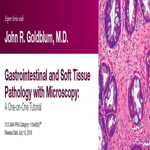
2019 Expert Series with John R. Goldblum, M.D. Gastrointestinal and Soft Tissue Pathology with Microscopy A One-on-One Tutorial (CME VIDEOS)
Description:
2019 Expert Series with John R. Goldblum, M.D. Gastrointestinal and Soft Tissue Pathology with Microscopy A One-on-One Tutorial This CME Activity as a practical, clinically focused review of diagnostic ultrasound featuring urgent scanning procedures and techniques. A mix of ob-gyn, abdominal, and vascular lectures provide a well-rounded review of clinical imaging techniques. Faculty share scanning tips, protocols, and pitfalls providing an overall balance to the activity.
This activity is designed to educate those who order, perform, and interpret clinical ultrasound studies. It should be particularly valuable for diagnostic imaging physicians, ob-gyn physicians, vascular surgeons, and referring physicians who utilize ultrasound to evaluate the clinical status of their patients.
Educational Symposia
Physicians: Educational Symposia is accredited by the Accreditation Council for Continuing Medical Education (ACCME) to provide continuing medical education for physicians.
Educational Symposia designates this enduring material for a maximum of 10.0 AMA PRA Category 1 Credit(s)TM. Physicians should claim only the credit commensurate with the extent of their participation in the activity.
All activity participants are required to take a written or online test in order to be awarded credit. All course participants will also have the opportunity to critically evaluate the program as it relates to practice relevance and educational objectives.
AMA PRA Category 1 Credit(s)TM for these programs may be claimed until July 14, 2022.
This CME activity was planned and produced by Educational Symposia, the leader in diagnostic imaging education since 1975.
This CME activity was planned and produced in accordance with the ACCME Essential Areas and Elements.
At the completion of this CME activity, subscribers should be able to:
- Describe the most common well-differentiated lipomatous tumors and the major diagnostic criteria for separating them from one another.
- Identify the most common morphologic patterns seen in soft tissue tumors.
- Apply the most useful immunohistochemical and molecular diagnostic techniques in the evaluation of soft tissue tumors.
- Discuss updates in the most recent WHO classification of renal tumors.
- Restate common and uncommon diagnostic pitfalls in bladder pathology.
- Explain how to reliably utilize both morphology and immunohistochemistry to differentiate germ cell tumors from mimics.
- Identify the histologic features of select cutaneous fibrohistiocytic tumors.
- Recognize cutaneous adnexal tumors that are associated with hereditary cancer predisposition syndromes.
- Describe the most useful ancillary diagnostic tests to aid in the diagnosis of challenging melanocytic lesions.
- Discuss current clinicopathologic diagnostic criteria for Barrett esophagus.
- Recognize the appearance of several medications that can be identified on routine histology.
- Explain the most common forms of gastritis with emphasis on the autoimmune form.
- Restate the natural history and disease biology of ductal carcinoma in situ.
- Recognize cytomorphologic pitfalls encountered in interpreting cerebrospinal fluid.
- Describe mediastinal anatomy and cytology sampling techniques.
- Discuss the appropriate use of p16 immunohistochemistry and HPV RNA in situ hybridization.
- Explain the TCGA molecular classification system of endometrial carcinoma.
- Review the interpretative challenges that attend MMR immunohistochemistry and understand the clinical implications of loss in tumors of the gynecologic tract.
No special educational preparation is required for this CME activity
Video Package:
1. Controversies in the Diagnosis of Barretts Esophagus and Barretts-Related Dysplasia
2. Microscopy – Esophageal Pathology With a Focus on Barretts Esophagus and Barretts-Related Dysplasia
3. Nomenclature of Colorectal Polyps Controversies and Confusion
4. Microscopy Colorectal Polyps Basic Issues Including Controversies in Nomenclature
5. The Trouble with Fat Diagnostic Issues with Well-Differentiated Lipomatous Tumors
6. Microscopy – Well-Differentiated Lipomatous Tumors
7. A Practical Approach to Mesenchymal Tumors of the GI Tract
8. Microscopy – Gastrointestinal Mesenchymal Tumors
9. Common Morphologic Patterns in Soft Tissue Tumors
10. Fibrohistiocytic Tumors of Soft Tissue with a Focus on Fibrohistiocytic Tumors of Intermediate Malignancy
11. Inflammatory Bowel Disease and IBD-related Dysplasia One Pathologists Perspective
CME Release Date 7/14/2019
CME Expiration Date 7/14/2022
Pay methods and How to get it
1. Pay please as per the methods on site See here if want get another way to pay click her CONTACT WITH ME
2- take screen shot when you pay and send to [email protected]
3. Once paid please provide mail id (which does not have registered) and Full name in One MSG to [email protected] like below
-
Pay with your local payment methods
KSA (Saudia arabia )
/and all Arabic gulf countries / YEMEN/ Egypt VISA Debt card Moneygram /Pak/ iran/ indian/ Bitcoin btc TRC 20 westen union /paypal

