Release Date : sept 2024
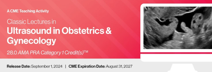
Classic Lectures in Ultrasound in Obstetrics & Gynecology – A CME Teaching Activity
This comprehensive CME teaching activity presents a series of classic lectures focused on ultrasound in obstetrics and gynecology. Covering both fundamental and advanced topics, the program includes in-depth reviews of the latest guidelines and innovations in fetal and obstetric imaging. The series features expert discussions on artificial intelligence in fetal cardiac imaging, first trimester ultrasound, and the diagnosis of complex conditions, among other critical areas. The faculty share tips, protocols, and advice on how to avoid pitfalls through actual clinical experiences.
Target Audience ▼
This CME teaching activity is designed to educate healthcare professionals involved in performing, ordering, and interpreting ultrasound examinations in obstetrics and gynecology. It is particularly valuable for obstetricians, gynecologists, maternal-fetal medicine physicians, radiologists, family medicine physicians, genetic counselors, and sonographers. Additionally, residents in obstetrics and gynecology, as well as fellows in maternal-fetal medicine and pediatric cardiology, will benefit from this program. The goal of this educational activity is to advance the level of practice in obstetric sonography and fetal echocardiography, enhancing diagnostic proficiency and patient care across these specialties.
Educational Objectives ▼
At the completion of this CME teaching activity, you should be able to:
- Review and differentiate between AIUM and ISUOG’s first trimester ultrasound guidelines.
- Apply artificial intelligence techniques in fetal cardiac imaging to improve diagnostic accuracy.
- Identify and evaluate the fetal brain, gastrointestinal tract, genitourinary system, heart, and skeletal system in first trimester ultrasounds.
- Diagnose common and rare syndromic conditions using first trimester ultrasound.
- Optimize imaging techniques for assessing fetal cardiac anatomy and anomalies, including great vessel abnormalities and cardiac tumors.
- Implement standardized approaches for diagnosing systemic venous malformations and congenital heart disease.
- Assess the role of genetic evaluation in the diagnosis of fetal anomalies and congenital heart disease.
- Integrate advanced ultrasound techniques, such as 3D/4D imaging, into routine practice for enhanced fetal assessment.
- Utilize ultrasound to manage ectopic pregnancies, placenta accreta, and various gynecological diagnoses, including adenomyosis and adnexal masses.
topics
- Adenomyosis: How to Make the Diagnosis with Ultrasound
Adnexal Masses in Pregnancy: Surgery or Follow-up?
Anomalies of the Great Vessels: Aortic Arch Abnormalities
Anomalies of the Great Vessels: Transposition of the Great Arteries
Approach to Diagnosis of Pulmonary Venous Malformations
Approach to Diagnosis of Systemic Venous Malformations
Artificial Intelligence in Fetal Cardiac Imaging
BI-RADS US Reviewonly at Medustudy.com
Bradyarrhythmias and Long QT Syndrome
Cardiac Imaging in Early Gestation
Cardiac Tumors
Cardiovascular Malformations of Twin-Twin Transfusion Syndrome
Commonly Missed GYN Diagnoses on US
Congenital Heart Disease in Multiple Pregnancies
Diagnosing Spina Bifida in the First Trimester
Ectopic Pregnancy: Here, There and Everywhere
Expanded Genetic Testing with Cell-Free DNA: What’s an Optimal Approach?
Fetal and Embryonic Sonographic Anatomy Before 11 Weeks
Fetal Ectopy and Tachyarrhythmias: Diagnosis & Management
First Trimester Ultrasound in the Diagnosis of Common and Rare Syndromic Conditions
First Trimester Ultrasound: The Fetal Brain
First Trimester Ultrasound: The Fetal Gastrointestinal Tract
First Trimester Ultrasound: The Fetal Genitourinary System
First Trimester Ultrasound: The Fetal Heart
First Trimester Ultrasound: The Fetal Skeletal System
Genetic Aspects of Congenital Heart Disease
Genetic Evaluation of Fetal Anomalies in the First Trimester
Guidelines for Performing a 2nd/3rd Trimester Level 1 Obstetrical Sonogram
Heterotaxy Syndrome
How to Incorporate Breast Elastography into Your Practice
Markers of Placenta Accreta In Early Gestation
National Guidelines for Fetal Echocardiography: What is Included?
Normal Fetal Cardiac Anatomy: The Cardiac Chambers
Obstetrical US “Aunt Minnie’s”
Optimizing Your Image in Fetal Cardiac Screening
O-RADS US in Daily Clinical Practice: Test Yourself! A Case Based Presentation
Oxygen for Fetal Intervention in Congenital Heart Disease: What is the Evidence?
Pregnancy Dating & Elements of Pregnancy Failure
Review of AIUM and ISUOG’s First Trimester Guidelines: How Do They Differ?
Systemic Fetal Venous Malformations: A Standardized Approach to Diagnosis
The Placenta & Umbilical Cord in Early Gestation
The Three-Vessel Trachea View
The Use of 3D in the 11-14 Weeks Ultrasound Examinations
The Use of 3D/4D Ultrasound in Fetal Cardiac Imaging
Tips & Tricks of Fetal Echocardiography
Ultrasound in the Diagnosis & Management of Ectopic Pregnancy
Ultrasound Signs of Aneuploidies at 11-14 Weeks
Update on Management of Pregnancies with Sjogren’s Antibodies
Update on Prenatal Diagnosis: CVS vs Amniocentesis
Update on US Evaluation of the Carotid Arteries
US Evaluation of Acute Pelvic Pain
US Evaluation of the Gravid Cervix: Measurements and More


