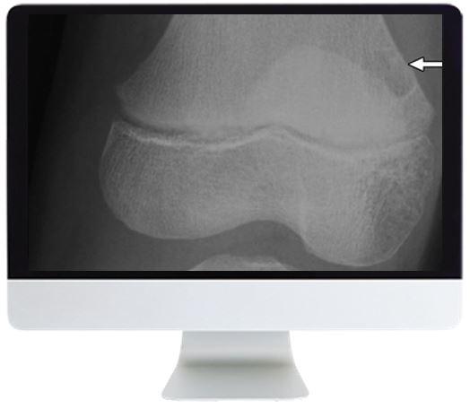
ARRS Clinical Case-Based Review of Musculoskeletal Imaging 2019
[
LEARNING OUTCOMES AND LECTURES
After completing this course, the learner should be able to:
- develop differential diagnoses for musculoskeletal imaging findings
- recognize the imaging and clinical features that allow for refinement of differential diagnosis, allowing for a more specific diagnosis
- recognize some commonly encountered imaging artifacts in musculoskeletal imaging and describe why they occur and the techniques to avoid them
- outline management decisions affecting a variety of commonly encountered clinical scenarios
Module 1
- Knee Trauma – Menisci—Daniel E. Wessell, MD, PhD
- Neoplasm—Jennifer L. Pierce, MD
- Trauma—Ambrose J. Huang, MD
- Inflammatory Conditions—F. Beaman
- Knee Trauma-Ligaments—Daniel E. Wessell, MD, PhD
- Rapid Fire Cases—Jonathan C. Baker, MD
Module 2
- Core—Ambrose J. Huang, MD
- Imaging Approach to Soft Tissue Masses—Jonathan C. Baker, MD
- Lower Extremity—Francesca Beaman, MD
- Characteristically Benign Soft Tissue Masses—Jonathan C. Baker, MD
- Upper Extremity—Hillary Warren Garner, MD
Module 3
- Rapid Fire Cases: Bone—Hillary Warren Garner, MD
- Imaging Approach: Muscle Disorders—Jonathan C. Baker, MD
- Rapid Fire Cases: Joint—Omer A. Awan, MD
- Imaging Approach: Tendon Disorders—Jonathan C. Baker, MD
- Rapid Fire Cases: Soft Tissue—James Derek Stensby, MD
]


